Your cornea is the thin outer layer of your eye that protects the eye from foreign debris. The shape of the cornea is also responsible for how well you can see clearly; the shape determines how light entering your eyes will focus in front of, behind or directly on the retina (the back of the eye).
5 Corneal Layers
- Epithelium (outer layer): eye protection and surface to absorb nutrients and oxygen from tears
- Bowman’s membrane (second layer): transparent collagen layer that can respond to injury by forming a scar
- Stroma (middle layer): thick collagen layer that provides elasticity and strength to form the cornea
- Descemet (inner layer): strong tissue that protects against injury and infection
- Endothelium (innermost layer): pumps excess fluid out of the stroma to keep the cornea clear
Types of Corneal Diseases
Corneal diseases can affect any layer of the cornea and ultimately result in distorted vision. Some of these diseases include:
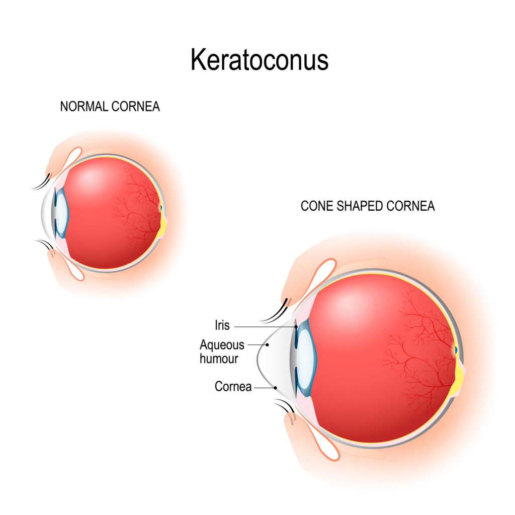
- Keratoconus: cone-shaped cornea due to corneal thinning
- Corneal dystrophies: accumulation of abnormal materials on the outer layer
- Keratitis: corneal inflammation
- Herpes of the eye: viral infection
- Shingles: corneal lesions or inflammation
- Pterygium: pink growth on the cornea
At Atwal Eye Care, our doctors offer different treatment options for corneal problems. A comprehensive dilated eye exam will help your doctor diagnose the issue and recommend the right course of action for your unique situation.
Keratoconus
Keratoconus is a condition that affects the cornea (the clear outer layer of your eye). Normally a round dome shape, if the corneal tissue begins to thin, it can begin to bulge out into a cone shape. Because the shape of the cornea becomes distorted, your eyesight also becomes distorted because light will not focus properly on the retina (back of the eye). This makes normal activities such as reading, watching TV, driving or working on a computer very difficult.
Who Gets Keratoconus
Many people are surprised to learn that keratoconus usually affects people who are in their late teenage years or early 20s. This is a condition that affects 1 in 2,000 of the general population. The condition is hereditary in some cases. It may also be caused by an enzyme imbalance in the cornea that causes the corneal tissue to weaken.
Symptoms of Keratoconus
Not all patients will experience keratoconus symptoms right away. In fact, it can progress over a 10-20 year period and result in symptoms such as:
- Vision distortion
- Blurry vision
- Light sensitivity
- Irritation
- Glare
- Ghosting
Keratoconus Treatment in Buffalo, NY
Luckily there are a variety of treatment options to help patients with keratoconus see more clearly. Depending on the severity of your condition our doctors may recommend:
- Prescription glasses
- Rigid contact lenses
- Corneal cross-linking (CXL)
- Intacs® corneal implants
- Corneal transplant in severe cases
Corneal Cross-linking for Keratoconus
If you suffer from keratoconus, your wait for progressive treatment is finally over. The FDA has approved corneal collagen cross-linking to strengthen weak corneas caused by keratoconus – and ultimately save vision. It may also reduce the need for corneal transplants for some patients.
We are excited to offer keratoconus patients corneal cross-linking using the Glaukos KXL System. This treatment is now widely covered by medical insurance.
How KXL Works
During this 2-step process, our doctors apply a riboflavin solution (a form of vitamin-B2) to the eye, followed by controlled ultraviolet (UV) light exposure for up to 30 minutes. This process increases the amount of collagen cross-links in the cornea, making the cornea stronger, more stable and able to hold its shape. A single treatment may be all that is needed to improve vision.
There are two types of KXL:
- Epithelium-off: the thin outer layer of the eye’s surface is removed and liquid riboflavin eye drops are applied
- Epithelium-on (transepithelial): liquid riboflavin eye drops are applied directly to the eye, without removing the thin outer layer of the eye’s surface
Only the epithelium-off method is currently FDA-approved in the U.S. The riboflavin eye drops used in this process are a combination of proprietary Photrexa and Photrexa Viscous solutions.
While not a cure for the disease, this keratoconus treatment has been effective in slowing the progression of the disease and helping preserve vision. Some patients may still need to wear prescription glasses or contact lenses to achieve their best vision possible.
Corneal Transplant
Because the cornea is so front and center on the eye, it is also prone to becoming damaged, diseased or scarred due to:
- Injuries
- Allergies
- Inflammation (keratitis)
- Infection
- Dry eye
- Thinning, causing a cone-shaped cornea (keratoconus)
- Cell deterioration (Fuchs’ dystrophy)
- Protein deposits (lattice dystrophy)
- Shingles
- Ocular herpes
- Pinkish growth (pterygium)
- Inward-facing eyelashes causing scarring (trichiasis)
- And a variety of other conditions
When this damage is severe enough, your vision will become greatly impacted. If glasses or contact lenses cannot restore your vision for daily tasks, your doctor may recommend a corneal transplant. This process works by removing all or part of the cornea and replacing it with healthy donor tissue from an eye bank.
At Atwal Eye Care, our doctors perform corneal transplants in Buffalo, NY. Depending on the severity of your problem, we may recommend:
- PK (Penetrating Keratoplasty): a full thickness corneal transplant to replace the entire cornea. Temporary sutures are necessary to keep the new cornea in place as the eye heals.
- DSAEK and DSEK (Descemet’s Stripping Automated Endothelial Keratoplasty and Descemet Stripping Endothelial Keratoplasty):two methods of partial thickness corneal transplants which replaces the damaged section of the innermost endothelium layer and the Descemet membrane. No sutures are required and most patients achieve a faster visual recovery.
- DMEK (Descemet Membrane Endothelial Keratoplasty): a partial thickness corneal transplant which involves removal of the Descemet membrane and endothelium which is then replaced with a very thin layer of donor tissue without additional stromal tissue.
If you are considering having a corneal transplant, make sure you are fully aware of the risks of surgery such as: rejection of the new tissue, infection and corneal swelling. Trusting your eyes to an experienced ophthalmologist will help reduce some of those risks. Contact our experienced team to learn more about your options.
Pterygium: Surfer’s Eye
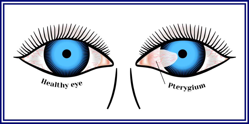
Pterygium is a noncancerous growth that looks like a raised white/pink tissue on the white of one or both eyes. It is often called surfer’s eye because it tends to affect people who often work or play outdoors, especially surfers.
While not typically a serious condition, pterygium can continue to grow until it covers the cornea, leading to distortion of the cornea (astigmatism) and blurred vision.
Surfer’s Eye Symptoms
- Blurred vision
- Redness
- Burning
- Itching
- Feeling of something foreign in the eye
Causes of Surfer’s Eye
Pterygium tends to affect people age 20 to 40 and it is more common in men. There is no single cause of pterygium; however, there are certain conditions that may increase your risk:
- Excessive exposure to UV light (being outdoors)
- Exposure to dust, wind, sand and other irritants
- Dry eye
- Not wearing sunglasses or hats outdoors
- Low humidity environments
Surfer’s Eye Treatment in Buffalo, NY
Medicated or lubricating eye drops can help ease irritation caused by pterygium. If the lesion begins to threaten your sight or makes you feel self-conscious, pterygium surgery to remove the growth may be recommended. To reduce the risk of recurrence, our doctors may perform an amniotic membrane transplant (AMT) graft where the former pterygium was located.
To learn more about corneal conditions, contact us today at 716-896-8831 to schedule an eye exam.